Lab Technique |
|
|
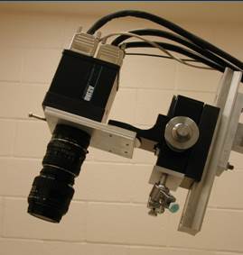
Intrinsic Signal Optical Imaging (ISOI) ISOI technique uses the intrinsic optical signal (hemodynamic signal), images the cortical activity with a highly-sensitive CCD camera. It is used to study the sub-millimeter functional structures (functional domains) and their contribution to the functions of the brain. This technique can achieve a very high spatial resolution (~0.1mm) and a relatively large area (up to 25mm). It is often combined with many other neural research methodologies (e.g. electrophysiology, histology, animal behavior). We use it to obtain cortical functional maps (e.g. Li Zhu et al. 2013); or quantitatively measure the cortical response to visual stimulation (e.g. Xu Han et al. 2016 ) |
|
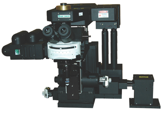
2-photon imaging We are currently developing 2-photon fluorescent imaging methods in studying cortical organization and functions of the primate brain. Two-photon imaging uses laser scanning for fluorescent activation, thus has high spatial resolution and deep penetration. With this technique we can monitor the activity of hundreds or thousands of neurons simultaneously. This technique is widely used in in vivo brain studies of small animals. Only several labs have reported its application in primate brain research. Recently we used this technique in studying the fine cortical structures in area V4 (Tang et al. 2020 ) |
|
Histology With optical imaging we can obtain functional information of the cortex. With histological methods, we can obtain anatomical information, which usually correlates with the functional organization. We use tracing methods to study the histological features of a specific type of neurons and their connections with distant functional domains identified with optical imaging. |
|
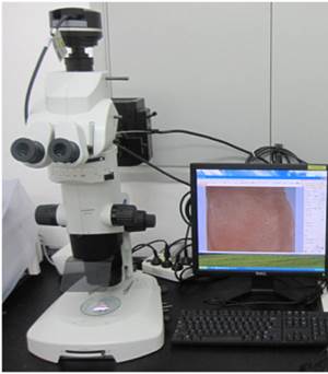
In vivo single-unit electrophysiology A classical technique used in studying neuronal function. We use it in combination with optical imaging (map – guided - recording) to study the receptive field properties of specific types of visual neurons (e.g. Li Zhu et al. 2013;Hu et al. 2018 ). |
|
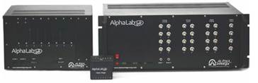
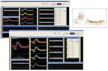
Multi-electrode array recording We recently started multi-electrode array recordings on monkey visual cortex. Arrays are 32- or 64-channel floating arrays and were implanted to specific cortical regions identified with optical imaging. With multi-neuron recording, we aim to know more about functional circuits for specific neuron types (see Ma et al. 2021 ). |
|
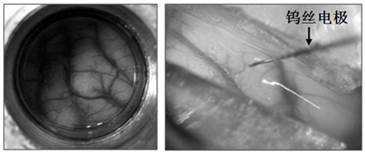
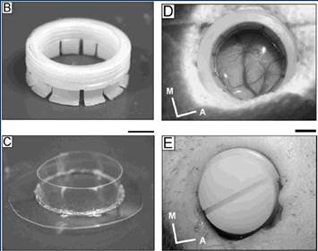
Chronic optical chamber We use chronic optical chambers for intrinsic signal optical imaging. The cortex is protected by artificial dura (panel C on the left) which is transparent for direct imaging of the cortex. Such chambers can be maintained and imaged for months to years. (image adapted from: Chen et al 2002, J Neurosci Method; for repeated imaging of cortical maps see Figure 4 in Li, Zhu et al. 2013 ) |
|
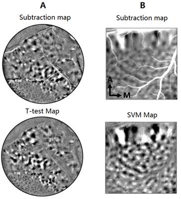
Image Analysis & Modeling We use different image analysis methods to extract useful information from weak signals in optical imaging data. Images on the left show the different signal-noise ratio for subtraction maps and t-test maps (A), and subtraction maps and SVM maps (B). (Figures are from Li, Zhu, et al. 2013 ,Figure S2 ;Chen et al. 2016 ,Figure S1). In temporal domain, we analyzed the temporal resolution of hemodynamic signals collected with optical imaging and found that such “slow” signals actually has a high temporal precision which can be used to track fast changing cortical dynamics (Lu et al. 2017). Based on this knowledge, we obtained single-trial timecourse of V1 optical responses to binocular rivalry stimulation (Xu Han et al. 2016 ). We are also collaborating with Dr. Malte Rasch and Dr. Si Wu on modeling our optical imaging data. In a recent study we successfully predicted V1 and V2 responses to moving random dots based on population response models (Rasch et al. 2013). |