Research Progress |
|
|
Shape-from-motion: the role of area V2
Human and non-human primates are good at identifying an object based on its motion, a task that is believed to be carried out by the ventral visual pathway. However, the neural mechanisms underlying such ability remains unclear. We trained macaque monkeys to do orientation discrimination for motion-boundaries (MB) and recorded neuronal response in area V2 with microelectrode arrays. We found 10.9% of V2 neurons exhibited robust orientation-selectivity to MBs, and their responses correlated with monkeys’ orientation-discrimination performances. Furthermore, the responses of V2 direction-selective neurons recorded at the same time showed correlated activity with MB neurons for particular MB stimuli, suggesting that these motion-sensitive neurons made specific functional contributions to MB discrimination tasks. Our findings support the view that V2 plays a critical role in MB analysis and may achieve this through a neural circuit within area V2. (details in: Ma et al. 2021 eLife. PDF ) |
|
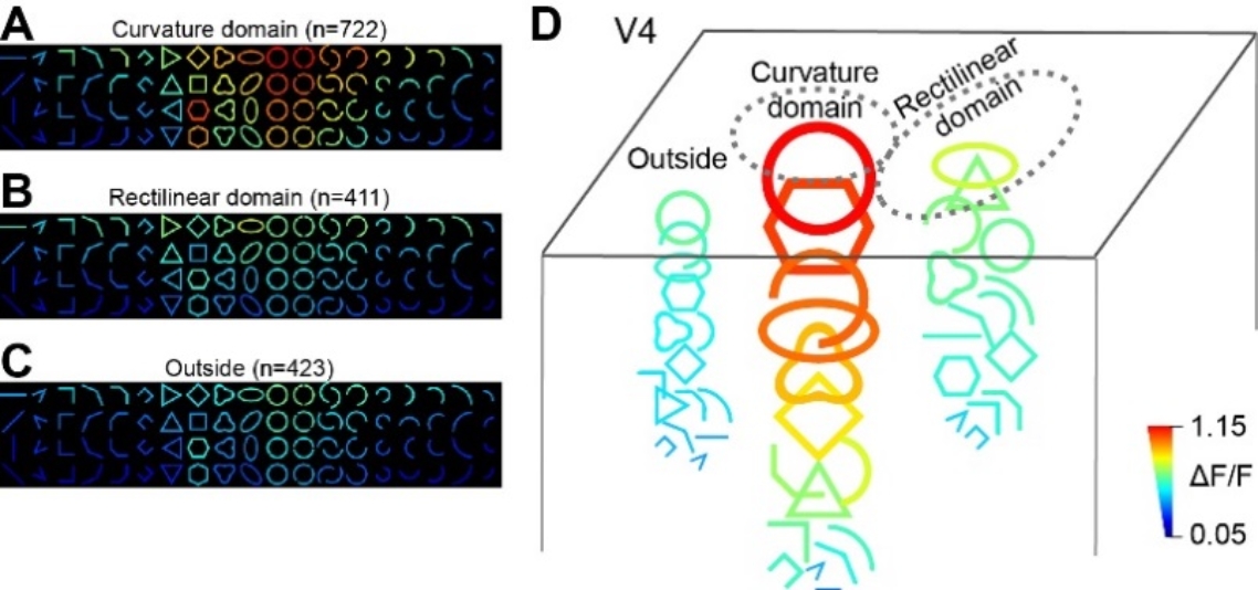
Seeing shape: curvature domains in visual area V4 Neurons in primate V4 exhibit various types of selectivity for contour shapes, including curves, angles, and simple shapes. How are these neurons organized in V4 remains unclear. Using intrinsic signal optical imaging and two-photon calcium imaging, we observed submillimeter functional domains in V4 that contained neurons preferring curved contours over rectilinear ones. These curvature domains had similar sizes and response amplitudes as orientation domains but tended to separate from these regions. Within the curvature domains, neurons that preferred circles or curve orientations clustered further into finer scale subdomains. Nevertheless, individual neurons also had a wide range of contour selectivity, and neighboring neurons exhibited a substantial diversity in shape tuning besides their common shape preferences. In strong contrast to V4, V1 and V2 did not have such contour-shape-related domains. These findings highlight the importance and complexity of curvature processing in visual object recognition and the key functional role of V4 in this process. (details in: Tang et al. 2020 eLife. PDF ) |
|
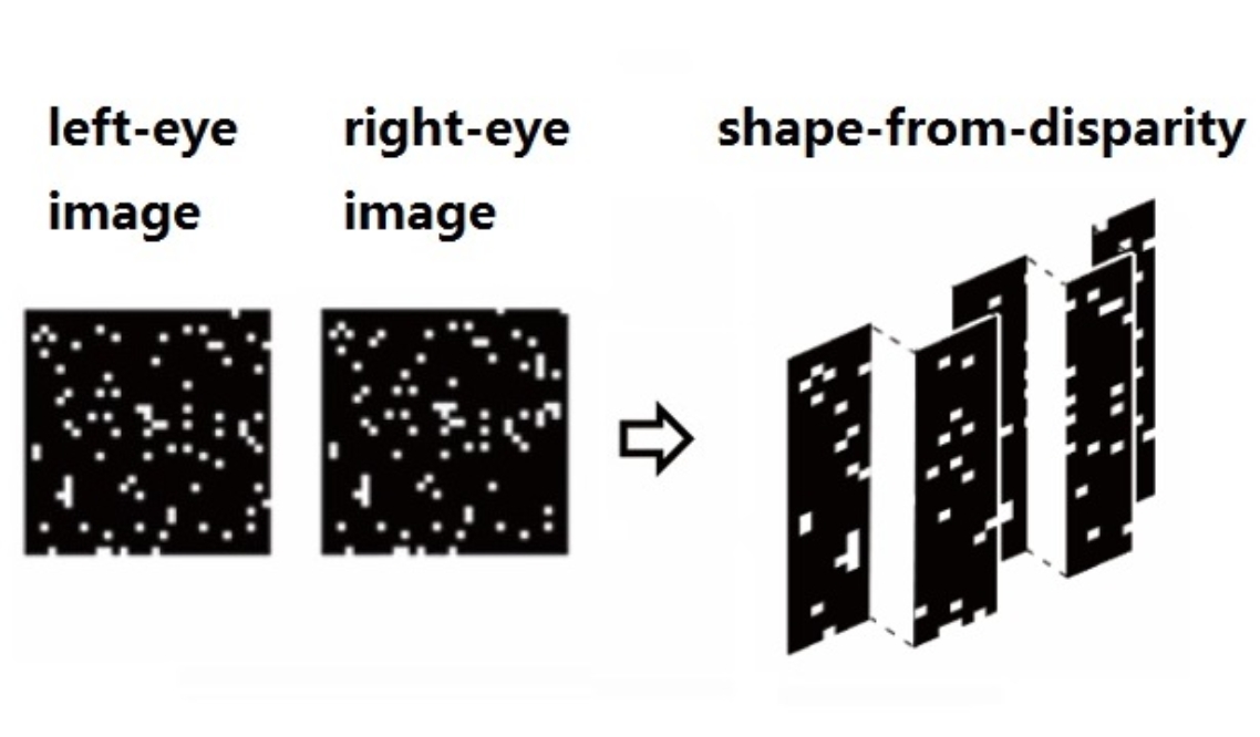
3D vision: an orientation map for disparity edges Binocular disparity is the small differences in the visual field seen by the two eyes. It is an important source of 3D perception, and is the basis for creating 3D movies and virtual reality. In our recent study, we created 3D contour edges purely based on binocular disparity in random dots (e.g. vertical strips on the left figure). We found that, there is an orientation map in monkey V4 activated by such disparity edges. This map is consistent with the orientation map obtained with regular luminance-defined edges, indicating a cue-invariant edge representation in this area. In contrast, such a map is much weaker in V2 and totally absent in V1. These findings reveal a hierarchical processing of 3D shape along the ventral pathway and the important role that V4 plays in shape-from-disparity detection. This is also the first functional architecture discovered in the primate visual system for shape-from-disparity. (details in: Fang et al. 2019 Cerebral Cortex. PDF ) |
|
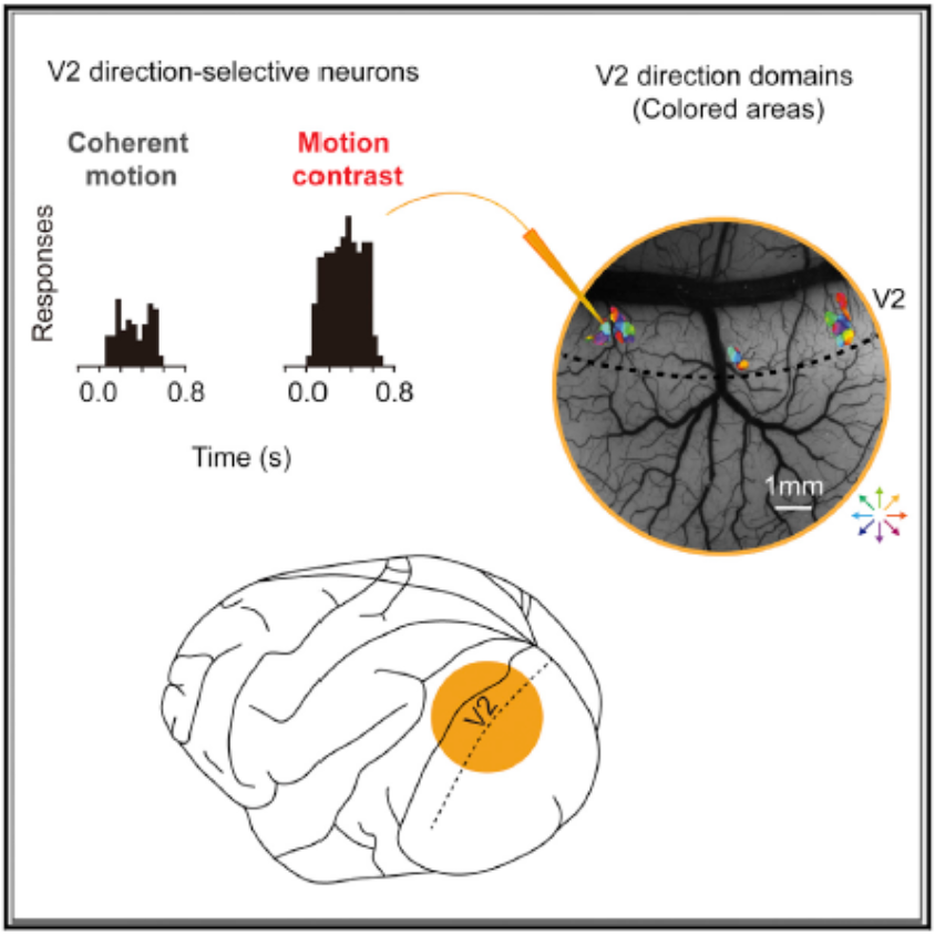
Visual motion: V2 detects motion contrast In the primate visual system, direction-selective (DS) neurons are critical for visual motion perception. While DS neurons in the dorsal visual pathway have been well characterized, the response properties of DS neurons in other major visual areas are largely unexplored. Recent optical imaging studies in monkey visual cortex area 2 (V2) revealed clusters of DS neurons. This imaging method facilitates targeted recordings from these neurons. Using optical imaging and single-cell recording, we characterized detailed response properties of DS neurons in macaque V2. Compared with DS neurons in the dorsal areas (e.g., MT), V2 DS neurons have a smaller receptive field and a stronger antagonistic surround. They do not code speed or plaid motion but are sensitive to motion contrast (top left figure). Our results suggest that V2 DS neurons can provide a saliency map (bottom left figure) of motion contours, where motion contrast is strongest, and thus play an important role in figure-ground segregation. The clusters of V2 DS neurons are likely specialized functional systems for detecting motion contrast. (details in: Hu et al. 2018 Cell Reports. PDF ) |
|
Neural mechanisms underlying binocular rivalry Binocular rivalry (BR) is a visual phenomenon where viewing two different monocular images gives rise to alternating percepts of the two monocular images. BR has been used as a tool in studying neural correlates of awareness and consciousness. It is unclear how much the neural activities in area V1 contribute to rivalry perception and whether top-down attention is necessary for rivalry to occur. To investigate this, we examined whether V1 neural activities alternate like the typical rivalry perception when top-down attention is deprived in anesthetized animals. Interestingly, in such a condition, V1 activity also exhibits left- and right-eye dominance alternation, just like what we would expect in awake animals. (details in: Xu, Han et al. 2016 The Journal of Neuroscience. PDF ) |
|
Motion boundary detection in the brain One type of visual stimulus we used to test cortical visual response. Such stimuli contain 2nd order contour information (horizontal or vertical lines) that are induced purely by relative motion of the random dots. (Note that random dots in two movies have the same diagonal motion.) We found some neurons in visual area V2 can “see” such horizontal and vertical lines just like they can “see” the real ones (e.g. the horizontal and vertical borders of these square figures). (details in Chen et al. 2016 Cerebral Cortex, PDF ) |
|
A motion map revealed in the ventral visual pathway In humans and other primates, visual information is processed in a parallel fashion, along different visual pathways. In the classical view, visual motion information is processed in the dorsal visual pathway, not the ventral visual pathway. A recent study in our lab showed that, in contrast to the classical view, motion information is processed in both the dorsal and ventral visual pathways, and possibly serves different purposes. Using intrinsic signal optical imaging, we mapped the direction of motion responses over large fields of view in V1, V2, and V4 in anesthetized monkeys. We found that V4 contains direction-preferring domains (~0.3mm diameter) that are preferentially activated by visual stimuli moving in one direction. This is the first demonstration of a motion organization in the ventral visual pathway. (details in Li, Zhu et al. 2013 Neuron, PDF ) |
|
Brain images of a moving dot It was believed that hemodynamic signals, like ISOI or fMRI BOLD are too slow to reflect temporal dynamics of neuronal activity. Here we used intrinsic signal optical imaging to track cortical activation in monkey V1, evoked by a small moving dot on a computer screen. The left video clip shows that the activated hemodynamic signals exhibited rich temporal dynamic information, including a “negative – positive - negative” tri-phasic signature, with different spatial distributions. Based on the temporal delays among different pixels, we were able to precisely measure the relative activation times of these pixels. The temporal precision can achieve<100ms. Thus hemodynamic signals also can be used in studies require high temporal precision. The video on the left was collected from an anesthetized monkey and averaged over 20 times of monocular visual stimulation. The moving dot had a size of 0.1 degree and moving speed was 2 degree/sec. Imaging frame rate was 8Hz and imaging duration was 10 sec. The imaged V1 region is about 2 cm diameter. This work was performed by Haidong Lu in Anna Roe’s lab (details in Lu, et al. 2017 Neuroimage, PDF ). |


